Open source

You are here

Open source software license

VTK

The Visualization Toolkit (VTK) is an open-source, freely available software system for 3D computer graphics, image processing and visualization. Kitware, whose team created and continues to extend the toolkit, offers professional support and consulting services for VTK. VTK supports a wide variety of visualization algorithms including: scalar, vector, tensor, texture, and volumetric methods; and advanced modeling techniques such as: implicit modeling, polygon reduction, mesh smoothing, cutting, contouring, and Delaunay triangulation. VTK has an extensive information visualization framework, has a suite of 3D interaction widgets, supports parallel processing, and integrates with various databases on GUI toolkits such as Qt and Tk.

Release Date:
Status:
Availability:
Data type:
Techniques:
Software:
Technology:
Platform:
Requirements:

Project development

Institution: Kitware, Inc.

Jalview

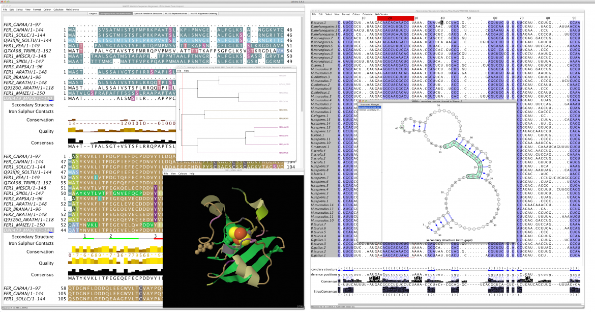
Jalview is a free program for protein and nucleic acid multiple sequence alignment editing, visualisation and analysis. Use it to view and edit sequence alignments, analyse them with phylogenetic trees and principal components analysis (PCA) plots and explore molecular structures and annotation. Jalview has built in DNA, RNA and protein sequence and structure visualisation and analysis capabilities. It uses Jmol to view 3D structures, and VARNA to display RNA secondary structure. The Jalview Desktop can also connect with databases and analysis services, and provides a graphical interface to the alignment and analysis services provided by the JavA Bioinformatics Analysis Web Services framework.  From Jalview 2.8.2, the Jalview desktop also interfaces with the Chimera molecular visualisation system to provide powerful 3D graphics tightly coupled to Jalview's functions.

Release Date:
Status:
Availability:
Data type:
Techniques:
Software:
Technology:
Platform:
Requirements:

Project development

Institution: University of Dundee

StratomeX


StratomeX is a tool designed for the discovery and characterization of disease subtypes based on genomic and clinical data of patient cohorts with hundreds of patients. The tool provides an interactive interface that enables analyst to quickly explore complex and heterogeneous datasets. 

Stratomex is part of Caleydo, an open source visual analysis framework targeted at biomolecular data.

Release Date:
Status:
Availability:
Data type:
Techniques:
Software:
Technology:
Platform:
Requirements:

Project development

Institution: Harvard

NetBioV

NetBioV (network biology visualization) is an R package that provides many easy to use functions, layout styles and color schemes to visualize biological networks

Release Date:
Status:
Availability:
Data type:
Techniques:
Software:
Technology:
Platform:
Requirements:

Project development

Institution: Computational Biology and Machine Learning

Gitools

Gitools is a desktop application for analysis and visualization of multidimensional data using interactive heat-maps.

Release Date:
Status:
Availability:
Data type:
Techniques:
Software:
Technology:
Platform:
Requirements:

Project development

Institution: University Pompeu Fabra, Barcelona

MembraneEditor

The CELLmicrocosmos 2.2 MembraneEditor can be used to model membranes based on lipid and protein files in PDB format. It is possible to interactively generate structural and compositional heterogeneous membrane patches as well as vesicles. The created structures can be used for visualization purposes, structural analysis or as a base for molecular simulations. Windows, Mac OS X as well as Linux are supported. Requirement: Java 7+ has to be installed beforehand. For semi-automatic placement of proteins, the OPM and PDB_TM database are used. A direct connection to the PDB database for downloading PDB files is also integrated. The PDB export module provides many options, enabling compatibility to all PDB viewers to the best of our knowledge.

Introduction videos you will find here: http://www.cellmicrocosmos.org/index.php/videos

Release Date:
Status:
Availability:
Data type:
Techniques:
Software:
Technology:
Platform:
Requirements:

Project development

Institution: University of Konstanz

LEVER

LEVER 3-D is an application that enables the quantitative analysis of multichannel 5-D (x, y, z, t, channel) and large montage confocal fluorescence microscopy images. The image sequences show stem cells together with blood vessels, enabling quantification of the dynamic behaviors of stem cells in relation to their vascular niche, with applications in developmental and cancer biology. LEVER 3-D automatically segments, tracks, and lineages the image sequence data and then allows the user to view and edit the results of automated algorithms in a stereoscopic 3-D window while simultaneously viewing the stem cell lineage tree in a 2-D window. Using the GPU to store and render the image sequence data enables a hybrid computational approach. An inference-based approach utilizing user-provided edits to automatically correct related mistakes executes interactively on the system CPU while the GPU handles 3-D visualization tasks. Conclusions: By exploiting commodity computer gaming hardware, we have developed an application that can be run in the laboratory to facilitate rapid iteration through biological experiments. There is a pressing need for visualization and analysis tools for 5-D live cell image data. This tool is the first to combine all of these aspects, leveraging the synergies obtained by utilizing validation information from stereo visualization to improve the low level image processing tasks.

Release Date:
Status:
Availability:
Data type:
Techniques:
Software:
Technology:
Platform:
Requirements:

Project development

Institution: Drexel University

MIMTool


Background: To understand protein function, it is important to study protein- protein interaction networks. These networks can be represented in network diagrams called protein interaction maps that can lead to better understanding by visualization. We address the problem of drawing of protein interactions in Kohn's Molecular Interaction Map (MIM) notation. Even though there are some existing tools for graphical visualization of protein interactions in general, there is no tool that can draw protein interactions with MIM notation with full support. Results: MIMTool was developed for drawing protein interaction maps in Kohn's MIM notation. MIMTool was developed using the Qt toolkit libraries and introduces several unique features such as full interactivity, object dragging, ability to export files in MIMML, SBML and line drawing with automatic bending and crossover minimization, which are not available in other diagram editors. MIMTool also has a unique orthogonal edge drawing method that is both easy and more flexible than other orthogonal drawing methods present in other interaction drawing tools. Conclusions: MIMTool facilitates faster drawing, updating and exchanging of MIMs. Among its several features, it also includes a semi-automatic drawing algorithm that makes use of shortest path algorithm for constructing lines with small number of bends and crossings. MIMTool contributes a much needed software tool that was missing and will facilitate wider adoption of Kohn's MIM notation.

Release Date:
Status:
Availability:
Data type:
Techniques:
Software:
Technology:
Platform:
Requirements:

Project development

Institution: Bogazici University, Istanbul; Boston University, Boston; National Lab for Cancer Research, National Cancer Inst., NIH, Frederick

This work was supported by the Scientific and  Technological Research Council of Turkey (TUBITAK) under the grant number 107T382 and in part with Federal funds from the National Cancer Institute, National Institutes of Health, under contract number HHSN261200800001E. The content of this publication does not necessarily reflect the views or policies of the Department of Health and Human Services, nor does mention of trade names, commercial products, or organizations imply endorsement by the U.S. Government. It was also supported in part by the Intramural Research Program of the NIH, National Cancer Institute, Center for Cancer Research.
 

inPHAP

inPHAP is an interactive visualization tool for genotype and phased haplotype data. inPHAP features a variety of interaction possibilities such as zooming, sorting, filtering and aggregation of rows in order to explore patterns hidden in large genetic data sets. As a proof of concept, we apply inPHAP to the phased haplotype data set of Phase 1 of the 1000 Genomes Project. Thereby, inPHAP's ability to show genetic variations on the population as well as on the individuals level is demonstrated for several disease related loci.

Release Date:
Status:
Availability:
Data type:
Techniques:
Software:
Technology:
Platform:
Requirements:

Project development

Institution: University of Tübingen

Deutsche Forschungsgemeinschaft and Open Access Publishing Fund of Tübingen University supported this project.

VIPER

VIPER (Visual Pedigree Explorer) is a tool for exploring large complex animal pedigrees and their associated genotype data. The tool combines a novel, space-efficient visualisation of the pedigree structure with an inheritance-checking algorithm. This allows users to explore the apparent errors within the genotype data in the full context of the family and pedigree structure. Ultimately, the aim is to develop an interactive software application that will allow users to identify, confirm and then remove errors from the pedigree structure and scored genotypes. 

Release Date:
Status:
Availability:
Data type:
Techniques:
Software:
Technology:
Platform:
Requirements:

Project development

Institution: The Roslin Institute University of Edinburgh, School of Computing Edinburgh Napier University

Pages